When a patient's mouth lacks bone as well as teeth, bone grafting, or the addition of bone, is required before an implant can be placed.
Bone grafting is where the jawbone is built up to accommodate a dental implant or other restorative device. Bone grafting is a common procedure that is used frequently for dental implants and other periodontal procedures. The bone used to graft is taken from a sample from the patient. Many times, the bone is taken from another area of the mouth when drilling takes place. The bone fragments are suctioned from the mouth and used for the graft. Cadaver bone fragments are also used. They are harvested by bone banks and are a very safe source for bone donation.
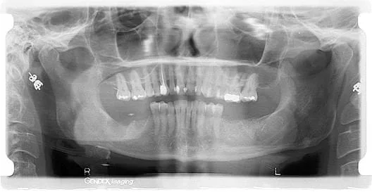
In the following Panoramic radiograph you can see the effect and importance of extraction site socket preservation. Notice how the area on the patient 's lower right is dark and uneven where there is loss of tooth structure and absence of bone following the extraction of two teeth. Now, notice on the patient's lower left where two teeth were extracted how the extraction sites are full and level with bone grafting material that will generate formation of natural bone in the patient's lower jaw. As a result, the area on the patient's lower left, once properly healed, can be restored with the placement of dental implants and crowns.
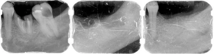
The photos below, in chronological order, demonstrate the process of extraction site socket preservation. The first radiograph reveals two deeply decayed teeth that can not be restored and must be extracted. The following radiograph was taken immediately after the teeth were removed, if the extraction sites were left this way without bone grafting the result would be an uneven ridge with approximately 60% loss of bone height in this area of the lower jaw. The final radiograph was taken after the bone grafting material was placed and the extraction sockets have now been preserved for future restoration.